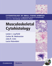
Musculoskeletal Cytohistology Hardback with CD-ROM
Each volume in this richly illustrated series, sponsored by the Papanicolaou Society of Cytopathology, provides an organ-based approach to the cytological and histological diagnosis of small tissue samples. Benign, pre-malignant and malignant entities are presented in a well-organized and standardized format, with high-resolution color photomicrographs, tables, tabulated specific morphologic criteria and appropriate ancillary testing algorithms. Example vignettes allow the reader to assimilate the diagnostic principles in a case-based format. This volume describes the cytologic, small core biopsy, immunohistochemical and molecular diagnostic features of musculoskeletal lesions. It provides the cytopathologist and histopathologist with a comprehensive survey of diagnostic approaches and diagnostic features for benign and malignant lesions. Each chapter contains a detailed summary of differential diagnostic features, supported by high-quality images and case studies. With over 500 printed photomicrographs and a CD-ROM offering all images in a downloadable format, this is an important resource for practising pathologists and residents in pathology.
- Richly illustrated with over 500 high-quality colour images; the print edition includes a CD-ROM containing all images in a downloadable format
- Case vignettes illustrate how to approach the cytologic diagnosis and histologic diagnosis based on mini core biopsies for a wide variety of musculoskeletal lesions
- Integrates modern diagnostic techniques with traditional cytologic and histopathologic analysis
Product details
March 2012Mixed media product
9781107014053
326 pages
283 × 225 × 19 mm
1.4kg
37 b/w illus. 577 colour illus. 17 tables
Available
Table of Contents
- Preface
- 1. Principles and practice for biopsy diagnosis and management of musculoskeletal lesions
- 2. Ancillary techniques useful in the evaluation and diagnosis of bone and soft tissue neoplasms
- 3. Spindle cell tumors of bone and soft tissue in infants and children
- 4. Spindle cell tumors of the musculoskeletal system characteristically occurring in adults
- 5. Giant cell tumors of the musculoskeletal system
- 6. Myxoid lesions of bone and soft tissue
- 7. Lipomatous tumors
- 8. Vascular tumors of bone and soft tissue
- 9. Pleomorphic sarcomas of bone and soft tissue
- 10. Osseous tumors of bone and soft tissue
- 11. Cartilaginous neoplasms of bone and soft tissue
- 12. Small round cell neoplasms of bone and soft tissue
- 13. Epithelioid and polygonal cell tumors of bone and soft tissue
- 14. Cystic lesions of bone and soft tissue
- Index.

