
Scanning Electron Microscopy for the Life Sciences
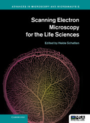
Scanning Electron Microscopy for the Life Sciences
CAD$195.95
Recent developments in scanning electron microscopy (SEM) have resulted in a wealth of new applications for cell and molecular biology, as well as related biological disciplines. It is now possible to analyze macromolecular complexes within their three-dimensional cellular microenvironment in near native states at high resolution, and to identify specific molecules and their structural and molecular interactions. New approaches include cryo-SEM applications and environmental SEM (ESEM), staining techniques and processing applications combining embedding and resin-extraction for imaging with high resolution SEM, and advances in immuno-labeling. New developments include helium ion microscopy, automated block-face imaging combined with serial sectioning inside an SEM chamber, and Focused Ion Beam Milling (FIB) combined with block-face SEM. With chapters written by experts, this guide gives an overview of SEM and sample processing for SEM, and highlights several advances in cell and molecular biology that greatly benefited from using conventional, cryo, immuno, and high-resolution SEM.
- Highlights novel applications of SEM in cell and molecular biology, covering cryo, immuno and high resolution SEM
- Instrumentation, sample preparation and imaging techniques are provided
- Dissemination of specific knowledge based on individual experience with emphasis on benefits and pitfalls
Reviews & endorsements
"This book provides not only basic information that will be useful to those new to microscopy, but also tips from experienced users that will surely aid other experienced microscopists … Recommended."
L. M. Baird, Choice
"[This book] is interesting for both beginners and advanced users. It is divided into fourteen chapters written by thirty-six contributors from nine different countries, which underlines its broad scope."
Jean-Marie Volland, Marine Ecology
Product details
January 2013Hardback
9780521195997
298 pages
244 × 170 × 17 mm
0.68kg
90 b/w illus. 39 colour illus.
Available


Table of Contents
- 1. The role of scanning electron microscopy in cell and molecular biology: SEM basics, past accomplishments and new frontiers Heide Schatten
- 2. Corrosion casting technique Jerzy Walocha, Jan A. Litwin and Adam J. Miodoński
- 3. Revealing the internal structure of cells in three dimensions with scanning electron microscopy Sol Sepsenwol
- 4. Mitochondria form continuous intracellular network-structures visualized with high-resolution field-emission scanning electron microscopy T. Naguro, H. Nakane and S. Inaga
- 5. Chapter on 3-D reconstruction of cell organelles using STEM tomography Paul Walther
- 6. High resolution labeling for correlative microscopy Ralph Albrecht, Daryl A. Meyer and O. E. Olorundare
- 7. The use of SEM to explore virus structure and trafficking Jens M. Holl and Elizabeth R. Wright
- 8. High resolution scanning electron microscopy of the nuclear surface in Herpes Simplex Virus 1 infected cells Peter Wild, Andres Kaech and Miriam S. Lucas
- 9. Scanning electron microscopy of chromosomes: structural and analytical investigations Elizabeth Schroeder-Reiter and Gerhard Wanner
- 10. A method to visualize the microarchitecture of glycoprotein matrices with scanning electron microscopy Giuseppe Familiari, Rosemarie Heyn, Luciano Petruzziello and Michela Relucenti
- 11. Scanning electron microscopy of cerebellar intrinsic circuits Orlando J. Castejón
- 12. Application of in vivo cryotechnique to living animal organs examined by scanning electron microscopy Shinichi Ohno, Nobuo Terada, Nobuhiko Ohno and Yasuhisa Fujii
- 13. SEM in dental research Vladimir Dusevich, Jennifer R. Melander and J. David Eick
- 14. SEM, teeth and palaeoanthropology: the secret of ancient human diets Alejandro Romero and Joaquín De Juan.