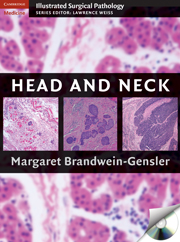
Head and Neck
Much of the diversity of head and neck diseases is due to the large number and broad function of the organs within this region. The surgical pathologist must become proficient in this subspecialty area in order to identify and categorize many different subtypes of lesions and diseases, including those affecting the thyroid and salivary glands. This book from the Cambridge Illustrated Surgical Pathology series comprehensively covers all of the methods utilized by pathologists to accurately diagnose diseases affecting all organs in the head and neck region. Coverage is not limited to findings from the light microscope but also includes other genetic, molecular, and immunologic diagnostic modalities, and the unique orientation allows the reader to follow the progression of disease states from incipient to advanced. This book is illustrated with more than 300 color photomicrographs and accompanied by a CD-ROM of all images in downloadable format.
- Illustrated with more than 300 color photomicrographs, accompanied by CD-ROM of all images
- Comprehensively covers all diseases and diagnostic modalities of the head and neck, including glands
- Unique orientation showcases diseases in various stages of development
Reviews & endorsements
"This is a practical, easy to read atlas of head and neck pathology. The bulleted outline presentation of the key histopathologic and differential diagnostic points makes it particularly easy to use."
--Doody's Review Service
Product details
October 2009Mixed media product
9780521879996
646 pages
285 × 220 × 45 mm
2.73kg
17 b/w illus. 538 colour illus. 33 tables
Available
Table of Contents
- 1. Sinonasal tract
- 2. Nasopharynx
- 3. Oral cavity
- 4. Larynx
- 5. Salivary glands
- 6. Thyroid and parathyroid glands
- 7. Jaws.

