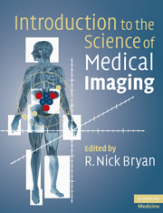
Introduction to the Science of Medical Imaging
Revolutionary advances in imaging technology that provide high resolution, 3-D, non-invasive imaging of biological subjects have made biomedical imaging an essential tool in clinical medicine and biomedical research. Key technological advances include MRI, positron emission tomography (PET) and multidetector X-ray CT scanners. Common to all contemporary imaging modalities is the creation of digital data and pictures. The evolution from analog to digital image data is driving the rapidly expanding field of digital image analysis. Scientists from numerous disciplines now require in-depth knowledge of these complex imaging modalities. Introduction to the Science of Medical Imaging presents scientific imaging principles, introduces the major biomedical imaging modalities, reviews the basics of human and computer image analysis and provides examples of major clinical and research applications. Written by one of the world's most innovative and highly respected neuroradiologists, Introduction to the Science of Medical Imaging is a landmark text on image acquisition and interpretation.
- Provides a survey of scientific imaging principles allowing the reader to put the myriad of methodological details into a common, unified framework
- Presents major points in verbal, pictorial and quantitative formats enabling readers to understand the increasingly critical link between images and numbers
- Clinical and research images used as methodological teaching tools – this fosters a deeper understanding of imaging technology as well as its practical applications
Reviews & endorsements
'This reviewer knows of no other source in medical imaging science that lays out the whole field so interestingly and with new twists in conveying information. It is a highly recommended purchase either for an individual or a department.' American Journal of Neuroradiology
'Very well written … Excellent images and diagrams … concise and up to date … a landmark text on the science of image interpretation. The authors manage to make a very complex subject very readable … relevant figures are exceptional …' BMA Medical Book Awards reviewer
'This is a most unusual and fascinating book. It blends science, philosophy and practical imaging in a very unusual way … As somebody closely involved in imaging I found it a fascinating read.' European Radiology
Product details
November 2009Paperback
9780521747622
334 pages
246 × 191 × 20 mm
0.91kg
159 b/w illus. 31 tables
Temporarily unavailable - available from TBC
Table of Contents
- Preface
- Part I. Introduction:
- 1. What is an image?
- 2. How to make an image
- 3. How to analyze an image
- Part II. Biomedical Images:
- 4. Nuclear medicine
- 5. X-ray: non-ionizing radiation
- 6. Ultrasound
- 7. Magnetic resonance imaging
- 8. Optical imaging
- Part III. Sample Labeling:
- 9. Contrast agents
- 10. Molecular labeling
- Part IV. Image Analysis:
- 11. Human observer
- 12. Digital image processing
- 13. Registration and atlas building
- 14. Statistical atlases
- Part V. Biomedical Applications:
- 15. Morphological imaging
- 16. Physiological imaging
- 17. Molecular imaging
- Appendices:
- 1. Linear systems
- 2. Transforms and k space
- 3. Probability, Bayesian statistics and information theory
- Index.


